Chronic Neuropathic Pain: Mechanisms and Common Subtypes
Chronic neuropathic pain is pain that persists or recurs for more than three months because a lesion or disease has affected the somatosensory nervous system—the network that carries information about touch, temperature, body position and potentially harmful stimuli.
The problem may arise in a peripheral nerve, nerve root, spinal cord or brain. Common examples include painful diabetic peripheral neuropathy, postherpetic neuralgia, painful radiculopathy, trigeminal neuralgia, pain following peripheral nerve injury, and central neuropathic pain after a stroke or spinal cord injury.
These conditions can share features such as burning, shooting or electric-shock-like pain, numbness, allodynia and hyperalgesia. However, symptoms alone do not prove that pain is neuropathic. Diagnosis requires evidence of a relevant lesion or disease and a pattern of pain that is anatomically consistent with the affected part of the nervous system.[1–4]
WHAT IS NEUROPATHIC PAIN?
The International Association for the Study of Pain defines neuropathic pain as “pain caused by a lesion or disease of the somatosensory nervous system.” The word dysfunction is not sufficient on its own: there should be a recognised neurological disease, a demonstrable lesion, or convincing clinical evidence of nervous-system injury.[1,2]
Neuropathic pain may be:
Peripheral, arising from a peripheral nerve, nerve root or sensory ganglion.
Central, arising from a lesion or disease affecting the spinal cord or brain.
Pain is classified as chronic when it persists or recurs for longer than three months.[3]
Neuropathic pain is different from nociceptive pain, which arises when a normally functioning sensory system detects actual or threatened injury to non-neural tissues such as joints, muscles or organs. It is also distinct from nociplastic pain, in which pain processing is altered without clear evidence of tissue injury sufficient to explain the pain or a lesion of the somatosensory system. These mechanisms are not mutually exclusive: more than one may contribute to an individual’s pain.[1,3]
Population studies suggest that pain with neuropathic characteristics affects approximately 7–10% of adults, although estimates vary because studies use different definitions, screening tools and diagnostic methods.[3,5]
WHAT CAN NEUROPATHIC PAIN FEEL LIKE?
Neuropathic pain may be continuous or intermittent. People commonly describe burning, shooting, stabbing, electric shocks, painful cold, squeezing or pins-and-needles sensations. Pain may occur spontaneously or be triggered by movement, temperature or touch.
Neuropathic pain can include both positive sensory symptoms, such as tingling, shocks or pain, and negative sensory symptoms, such as numbness or reduced ability to feel touch, temperature or pinprick. Pain and numbness can therefore occur in the same area.
Two commonly used terms are:
Allodynia: pain caused by a stimulus that would not normally be painful, such as clothing or light touch.
Hyperalgesia: an unusually strong response to a stimulus that is normally painful.
These findings are important clinical clues, but neither allodynia nor hyperalgesia identifies a specific mechanism by itself. Both can also occur in conditions that are not neuropathic.[1,6]
HOW IS NEUROPATHIC PAIN DIAGNOSED?
There is no single blood test, scan or questionnaire that diagnoses every form of neuropathic pain. Assessment usually considers four linked questions:
Is there a history of a neurological lesion or disease capable of causing the pain?
Is the pain located in a neuroanatomically plausible area—for example, the territory of an injured nerve or sensory pathway?
Are there compatible sensory changes on examination in the same area?
Is there a test that confirms the relevant lesion or disease?
The Neuropathic Pain Special Interest Group grading system classifies cases as possible, probable or definite neuropathic pain, according to the available clinical and investigative evidence.[2]
Depending on the suspected cause, investigations may include blood tests, nerve-conduction studies, electromyography, MRI, trigeminal reflex testing or skin biopsy for suspected small-fibre neuropathy. Screening questionnaires such as the DN4, LANSS or PainDETECT may help identify neuropathic features, but they do not replace clinical assessment.[2,4]
CORE MECHANISMS
Neuropathic pain is not produced by one universal pathway. Different mechanisms may occur at different sites, and their relative importance varies between conditions and between individuals.[6,7]
Abnormal electrical activity
An injured, compressed or demyelinated nerve may begin generating impulses without an appropriate external stimulus. This abnormal or ectopic activity can arise at an injured nerve ending, a neuroma, a compressed nerve root, a sensory ganglion or, in some central pain conditions, within the thalamus.
Changes in sodium, potassium and calcium channels can make sensory neurons more excitable and more likely to fire spontaneously. This can contribute to ongoing burning pain or sudden electric-shock-like attacks.[6,7]
Peripheral sensitisation and neuroimmune signalling
Nerve injury activates interactions between sensory neurons, supporting cells and immune cells. Signalling molecules released around an injured nerve can lower activation thresholds and increase abnormal firing.
This neuroimmune response is more complex than ordinary inflammation in a sprained joint or arthritic tissue. It may contribute to pain persistence, but inflammatory blood tests or cytokine testing are not currently used to diagnose neuropathic pain in routine clinical practice.[6,7]
Amplification within the spinal cord and brain
Abnormal incoming signals can change how sensory information is processed in the spinal cord and brain. Excitatory transmission may increase, inhibitory signalling may decrease, and spinal neurons may begin responding excessively to normal sensory input.
This helps explain why light touch can become painful or why pain may extend beyond the precise site of nerve injury. These changes are often described as central sensitisation, although that term refers to a physiological process rather than a diagnosis that can be confirmed from symptoms alone.[1,6]
Reduced or altered pain inhibition
The brain normally regulates incoming sensory information through descending pathways that can either suppress or facilitate pain. In some neuropathic pain conditions, inhibitory control becomes less effective or the balance shifts towards facilitation.
This does not mean that the brain has simply “failed to switch pain off.” Pain modulation is dynamic and can be influenced by the location of the lesion, ongoing sensory input, medication, sleep, stress and other aspects of health.[6]
Plasticity following loss of sensory input
A nerve lesion may reduce or distort the information reaching the spinal cord or brain. This loss of normal input—sometimes called deafferentation—can lead to abnormal activity and reorganisation within sensory networks.
Research has identified changes in brain activity and connectivity in groups of people with neuropathic pain. However, these findings are not sufficiently specific to diagnose an individual patient, and functional brain imaging is not currently recommended as a routine diagnostic test for neuropathic pain.[4,6]
COMMON SUBTYPES
PAINFUL DIABETIC POLYNEUROPATHY
Diabetic distal symmetrical polyneuropathy usually begins in the toes and feet, often on both sides, and may gradually extend upwards. Symptoms can include burning, tingling, electric pain, sensitivity to touch and numbness.
Not everyone with diabetic neuropathy experiences pain. International consensus estimates suggest that painful distal symmetrical polyneuropathy affects approximately 13–26% of people with diabetes, while up to half of people who have diabetic polyneuropathy may have few or no symptoms. Loss of protective sensation remains clinically important because injuries or ulcers may go unnoticed.[8]
Diabetic nerve injury is not caused by a single process. Metabolic stress, microvascular changes, oxidative stress, inflammation and other factors may affect both small and large nerve fibres. It is therefore more accurate than describing the condition simply as “small-fibre damage caused by high blood glucose.”[8]
POSTHERPETIC NEURALGIA
Postherpetic neuralgia is pain that continues or returns in the same area after shingles. It is commonly defined as pain persisting for at least three months after the shingles rash began, although definitions vary between studies.
The varicella-zoster virus can injure sensory ganglia, peripheral nerves and their connections within the spinal cord. Some people develop ongoing burning or aching pain, while others experience severe allodynia in which clothing or light touch becomes painful.
Approximately 10–18% of people who develop shingles experience postherpetic neuralgia, with the risk increasing substantially with age.[9]
PAINFUL RADICULOPATHY
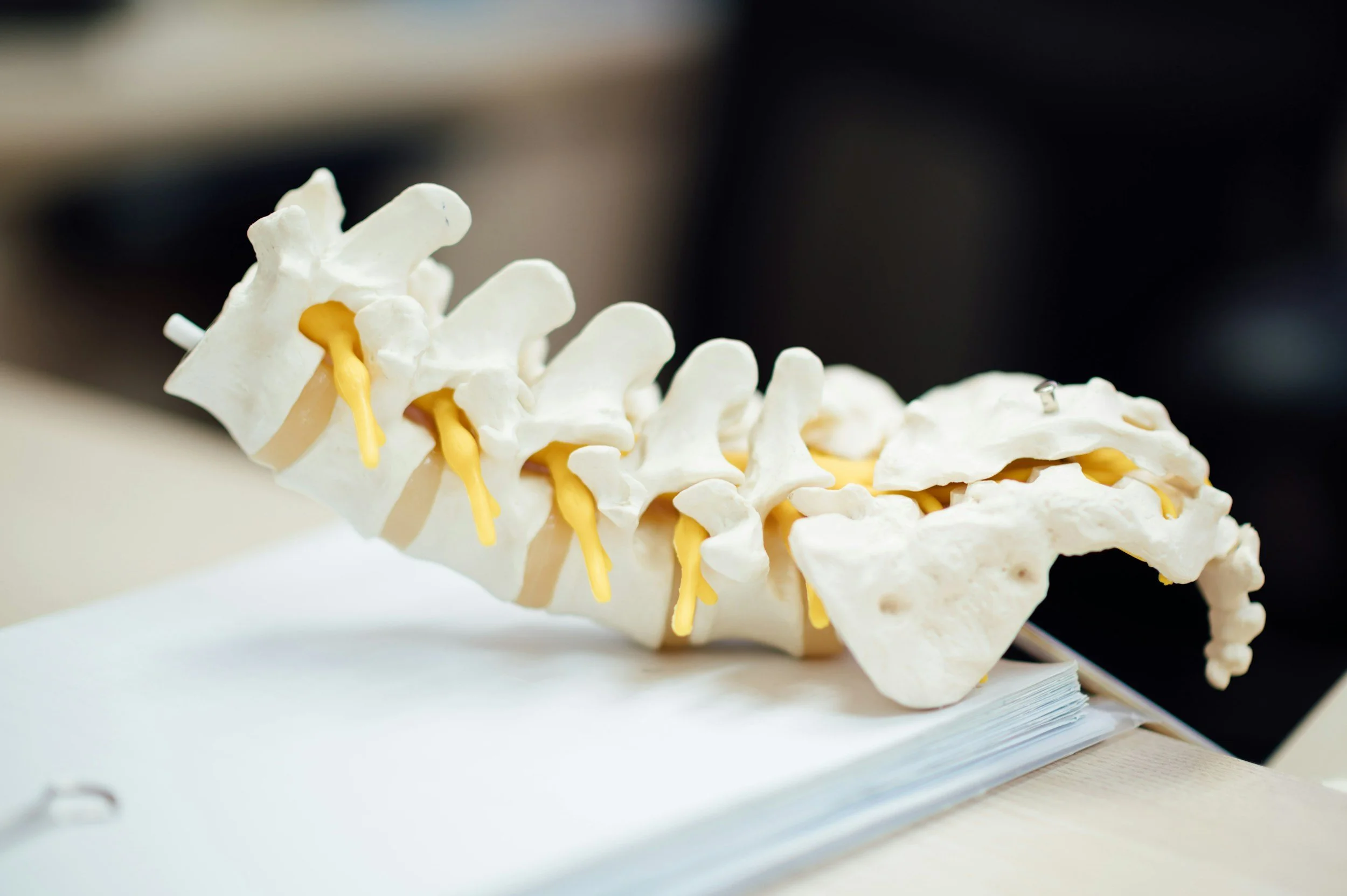
A radiculopathy results from a lesion or disease affecting a spinal nerve root. Causes include disc herniation, foraminal narrowing, spinal stenosis, inflammation or other structural disorders.
Pain may radiate along the affected nerve-root distribution and may be accompanied by numbness, altered reflexes or weakness. However, not every episode of “sciatica,” neck-to-arm pain or radiating back pain is neuropathic. Musculoskeletal referred pain can produce a similar distribution, and abnormalities on imaging do not automatically prove that a particular nerve root is causing the symptoms.
Clinical examination and correlation between the symptoms, neurological findings and imaging are therefore important.[2,3]
TRIGEMINAL NEURALGIA
Trigeminal neuralgia causes recurrent, brief attacks of severe, unilateral facial pain. The pain is typically electric-shock-like, shooting or stabbing and may be triggered by activities such as touching the face, chewing, speaking, washing or brushing the teeth. Individual attacks usually last from a fraction of a second to two minutes.[10]
In classical trigeminal neuralgia, a blood vessel commonly compresses the trigeminal nerve near the brainstem, producing focal demyelination and abnormal electrical “cross-talk” between nerve fibres. Secondary trigeminal neuralgia may occur because of multiple sclerosis or another structural lesion, while in some cases no cause is identified.
Trigeminal neuralgia has a distinctive assessment and treatment pathway. MRI is generally used to investigate possible secondary causes and neurovascular compression.[10,11]
PAINFUL PERIPHERAL NERVE INJURY
Trauma, surgery, entrapment or another focal injury may damage an individual peripheral nerve. Pain is usually located within the nerve’s sensory territory and may occur alongside numbness, weakness, a tender neuroma or marked sensitivity around a scar.
Not all persistent pain after surgery or trauma is neuropathic. A neuropathic component is more likely when there is evidence of nerve injury and compatible sensory abnormalities.[2,3]
CENTRAL NEUROPATHIC PAIN
Central neuropathic pain results from a lesion or disease affecting central somatosensory pathways. Recognised examples include:
Central post-stroke pain
Pain following spinal cord injury
Pain associated with multiple sclerosis
Pain following traumatic brain injury
The painful area and sensory abnormalities should be anatomically consistent with the central lesion. Not all pain experienced after a stroke, spinal cord injury or multiple sclerosis is neuropathic: musculoskeletal pain, spasticity-related pain, headache and other mechanisms may coexist.
Central pain is not limited to lesions of the thalamus. Injury anywhere along relevant spinal, brainstem, thalamic or thalamocortical sensory pathways may contribute.[3,6]
OTHER FORMS OF NEUROPATHIC PAIN
Other recognised causes include chemotherapy-induced peripheral neuropathy, HIV-associated neuropathy, alcohol-related or nutritional neuropathy, immune-mediated neuropathies and some inherited neurological disorders. Importantly, a neuropathy may produce numbness or weakness without pain, so “neuropathy” and “neuropathic pain” are not interchangeable terms.
Phantom limb pain also involves neuropathic mechanisms, but it should not be attributed solely to cortical reorganisation. Changes in the residual peripheral nerves, spinal cord and brain may all contribute.[6,7]

WHY CAN NEUROPATHIC PAIN PERSIST?
Neuropathic pain is sometimes described as a simple sequence of nerve injury, spinal “wind-up” and brain reorganisation. In practice, the process is neither this linear nor this uniform.
A lesion may generate ongoing abnormal impulses while simultaneously altering ion channels, immune signalling, spinal inhibition and central sensory processing. Several of these changes may operate in parallel. Some may diminish as the nervous system recovers, while others persist.
Nerve injury also does not inevitably cause chronic pain. Many people with comparable nerve lesions develop numbness without pain, while others develop severe spontaneous or evoked pain. Differences in the type and location of injury, metabolic health, genetics, immune responses and nervous-system plasticity may help explain this variation.[6,7]
As with all pain, the lived experience is influenced by biological, psychological and social factors. Poor sleep, fear, low mood, stress, reduced movement and social disruption can increase distress and disability. This does not mean the pain is imagined or that a demonstrable neurological lesion is unimportant. It means that effective care may need to address both the neurological disorder and its broader effects on the person’s life.[1]
TREATMENT IMPLICATIONS
Treatment should begin with identifying the underlying condition and determining whether further nerve injury can be prevented or treated. Depending on the cause, this might include optimising diabetes management and foot protection, correcting a nutritional deficiency, reviewing a potentially neurotoxic medication, treating an immune or infectious disorder, or assessing whether structural nerve compression requires specific intervention.
Medication
The 2025 NeuPSIG systematic review supports three medication classes as general first-line options for adult neuropathic pain:
Tricyclic antidepressants
Serotonin-noradrenaline reuptake inhibitors
α2δ calcium-channel ligands, including gabapentin and pregabalin
These recommendations apply across neuropathic pain generally and do not mean that every medication is suitable for every condition or every person. Choice depends on the pain subtype, other medical conditions, concurrent medication, kidney function, falls risk, potential adverse effects and individual treatment priorities.[12]
Topical lidocaine or capsaicin may be considered for selected forms of localised peripheral neuropathic pain. Availability, approved indications and funding vary between countries and products. Opioids are not recommended as routine first-line treatment and were placed among weakly recommended third-line options in the updated review.[12]
Trigeminal neuralgia is an important exception to the general medication pathway. Carbamazepine or oxcarbazepine is usually recommended as initial treatment, with surgical or other specialist options considered when medication is ineffective or poorly tolerated.[11]
No medication works for everyone, and benefit is often partial rather than complete. A time-limited therapeutic trial should therefore consider not only pain intensity but also sleep, activity, function, adverse effects and whether the person considers the overall benefit worthwhile.
Rehabilitation and whole-person care
Physical and occupational rehabilitation may help maintain movement, strength, balance, confidence and participation. Treatment can include functional goal-setting, pacing, graded activity, equipment or workplace modification, and condition-specific sensory rehabilitation.
Education, sleep treatment and psychological approaches can help people respond to pain flares, reduce fear and distress, and re-engage with valued activities. These treatments do not imply that the neurological pain is psychological. They address the consequences and modifiers of persistent pain while medical treatment addresses the underlying lesion and pain mechanisms.
Procedures, surgery, neuromodulation and infusion therapies have condition-specific roles for carefully selected patients. They should not be presented as interchangeable or generally effective for all neuropathic pain conditions.
KEY POINTS
Neuropathic pain is not simply severe pain, unexplained pain or pain that continues after an injury. It is a specific clinical category requiring a lesion or disease of the somatosensory nervous system and an anatomically plausible relationship between that lesion and the painful area.
Burning pain, electric shocks, allodynia and hyperalgesia can support the diagnosis, but do not establish it by themselves. Different neuropathic pain disorders share some biological mechanisms while also having distinct causes, assessments and treatment pathways.
Management is therefore individualised. Realistic goals commonly include reducing pain, improving sleep and function, preventing further nerve injury, and helping the person participate more fully in everyday life.
General information disclaimer
This article provides general educational information and is not a substitute for individual medical assessment, diagnosis or treatment. It does not recommend any specific investigation, medicine, procedure or other therapy. Treatment suitability varies according to the diagnosis, medical history, other medications and individual circumstances. Concerns about persistent pain should be discussed with an appropriately qualified health practitioner.
Last medically reviewed 15/08/2026
References
International Association for the Study of Pain. IASP Terminology: neuropathic pain, peripheral neuropathic pain, central neuropathic pain, allodynia, hyperalgesia and sensitisation. Accessed 15 August 2026.
Finnerup NB, Haroutounian S, Kamerman P, et al. Neuropathic pain: an updated grading system for research and clinical practice. Pain. 2016;157(8):1599–1606. doi:10.1097/j.pain.0000000000000492.
Scholz J, Finnerup NB, Attal N, et al. The IASP classification of chronic pain for ICD-11: chronic neuropathic pain. Pain. 2019;160(1):53–59. doi:10.1097/j.pain.0000000000001365.
Truini A, Aleksovska K, Anderson CC, et al. Joint European Academy of Neurology–European Pain Federation–Neuropathic Pain Special Interest Group guidelines on neuropathic pain assessment. Eur J Neurol. 2023;30(8):2177–2196. doi:10.1111/ene.15831.
van Hecke O, Austin SK, Khan RA, Smith BH, Torrance N. Neuropathic pain in the general population: a systematic review of epidemiological studies. Pain. 2014;155(4):654–662. doi:10.1016/j.pain.2013.11.013.
Finnerup NB, Kuner R, Jensen TS. Neuropathic pain: from mechanisms to treatment. Physiol Rev. 2021;101(1):259–301. doi:10.1152/physrev.00045.2019.
Costigan M, Scholz J, Woolf CJ. Neuropathic pain: a maladaptive response of the nervous system to damage. Annu Rev Neurosci. 2009;32:1–32. doi:10.1146/annurev.neuro.051508.135531.
Ziegler D, Tesfaye S, Spallone V, et al. Screening, diagnosis and management of diabetic sensorimotor polyneuropathy in clinical practice: international expert consensus recommendations. Diabetes Res Clin Pract. 2022;186:109063. doi:10.1016/j.diabres.2021.109063.
Centers for Disease Control and Prevention. Shingles symptoms and complications: postherpetic neuralgia. Updated 19 April 2024. Accessed 15 August 2026.
Headache Classification Committee of the International Headache Society. 13.1 Trigeminal neuralgia. In: International Classification of Headache Disorders, 3rd edition.
Bendtsen L, Zakrzewska JM, Abbott J, et al. European Academy of Neurology guideline on trigeminal neuralgia. Eur J Neurol. 2019;26(6):831–849. doi:10.1111/ene.13950.
Soliman N, Moisset X, Ferraro MC, et al. Pharmacotherapy and non-invasive neuromodulation for neuropathic pain: a systematic review and meta-analysis. Lancet Neurol. 2025;24(5):413–428. doi:10.1016/S1474-4422(25)00068-7.